1. Detection method of naringinase enzyme activity
1-1 Spectrophotometer method
The para nitrophenol spectrophotometric method uses p-nitrophenol- α-rhamnoside ( pNP- α- Rha ) and p-nitrophenol-β-D-glucopyranoside ( pNP- β-Glu) as substrate, for detecting the enzyme activity of the α-rhamnosidase and β-glucosidase. Li Ping used the p-nitrophenol spectrophotometer method, in the experiment of adding Aspergillus niger β-glucosidase into the solution of p-nitrophenol -β-glucoside ( pNPG ) for enzymatic hydrolysis, and the value of absorbance was measured at 400 nm ultraviolet wavelength. And on the basis of the value of absorbance, they calculates the enzyme activity of the β -glucosidase. This method has strong substrate specificity, but the standard substrate is expensive, and the enzyme activity of naringinase to hydrolyze glycosides in food cannot be determined, so this method is not suitable for the detection of enzyme activity in industrial large scale preparation of naringinase.
The principle of the Davis spectrophotometer method is that naringin can occur a chromogenic reaction reaction with 90% diethylene glycol under slightly alkaline conditions, and the yellow naringin chalcone production can be detected under ultraviolet light with a wavelength of 420 nm. According to the measured absorbance value , the remaining amount of the substrate naringin can be calculated, and then the naringinase activity can be obtained by calculation. Wang Weina et al. used an improved Davis method, took naringin as a substrate, and added naringinase produced by liquid fermentation of Aspergillus niger FFCC 848 for enzymatic hydrolysis. The absorbance was measured under 420 nm ultraviolet light, and then calculated the enzyme activity. Davis spectrophotometer method for measuring the enzyme activity is simple for operation, have a fast speed, so this method is widely used in eeal time detection of naringinase production by fermentation and activity of immobilized naringinase.
1-2 High performance liquid chromatography
The principle of high performance liquid chromatography ( HPLC) is that HPLC can accurately determine and quantitative analyze the substrate naringin, the intermediate product purunin and the final product naringenin during the entire hydrolysis process , and then calculate the enzyme activity. Chen Yuelong and others used HPLC to determine the retention time of naringin, arbutin, myricetin and their products, and achieved complete separation with a resolution greater than 1.5, and effectively monitored the effect of naringinase on multiple glycosides in hydrolysis process, at the same time, the enzyme activity was determined accurately. The HPLC method has the advantages of high accuracy and can avoid the interference of impurities, but compared with the Davis method, this method has a longer detection time and more complicated operation.
1-3 Improved determination method for naringinase enzyme activity
Using the improved Davis method: 0.8 mL of 0.8 g·L-1 naringin solution prepared with pH 4.5 buffer was added into the reaction system containing 40 mg of immobilized naringinase (or 0.2 mL of free naringinase). Place it in a 50 ℃ constant temperature water bath for 30 min. Take 0.1 mL of the reaction enzymatic hydrolysis solution and add 5 mL diethylene glycol (90% volume fraction) and 0.1 mL NaOH solution (4 mol·L-1) in tubes, and stand for 10 min after mixing them uniformly. Measure the absorbance of the mixed solution at 420 nm.
The definition of the activity of naringinase enzyme (U) : at a condition of pH 4.5, 50 ℃, 1 enzyme activity unit is the amount of enzyme required to degrade 1 μg naringin per minute. Enzyme activity ratio ( U·g-1 or U·mL-1) is defined as: at the condition of pH 4.5, 50 ℃, enzyme activity exhibited by 1 g immobilized naringinase (or 1 mL free naringinase). Relative enzyme activity is calculated according to the following formula :
Determination of standard curve: the concentration of mass of standard glycoside naringin solution are arranged 0, 0.4, 0.8, 1.2, 1.6 g · L-1, and then 0.1 mL solution was taken for reaction with 5 mL of diethylene glycol (90% , v / v) and 0.1 mL sodium hydroxide solution ( 4 mol·L-1). After the mixture was evenly mixed and stood for 10 min, the light absorption value of the mixed solution was measured at 420 nm, and the standard curve was drawn accordingly.
2. Preparation of naringinase enzyme solution
The Aspergillus niger FFCC uv-11 stored at 4 ℃ was transferred to the slant medium, and the mature spores were obtained by constant temperature culture at 30 ℃ for 96 h . Then, the spores were washed with sterile normal saline with a mass fraction of 0.9%. the value of absorbance of spore suspension is measured at 600 nm (OD600 value), with sterile saline its OD600 value was adjusted to 0.200. And then the spore suspension with the volume fraction of 10% inoculation amount to 100 mL of fermentation medium (250 mL conical flask), The obtained fermentation broth was centrifuged at 4 ℃ and 6000 r·min-1 for 10 min in a refrigerated centrifuge. Then through the suction filter to get supernatant fluid required naringin enzyme enzyme fluid. Chill it at 4 ℃ for later use.
3. Preparation of Chitosan Immobilized Naringinase enzyme
Take 3.0 g of chitosan, with 100 mL of volume fraction of 2% acetic acid solution was dissolved to prepare 0.3 g · L-1 chitosan colloidal solution, sonicated for 20 min to remove air bubbles and allowed it to fully dissolve. The chitosan colloidal solution was injected drop by drop with a needle into 0.5 g · L-1 NaOH condensate (1000 mL) to form a smooth pellets. Allow it to stand at room temperature for 3 h, and the pellets were repeatedly washed with distilled water to neutral, and the water was absorbed by a filter paper to obtain the chitosan microspheres. 0.4 g chitosan microspheres were weighed, 0.4 ml glutaraldehyde with 6% volume fraction was added, and the microspheres were repeatedly washed with distilled water until there was no glutaraldehyde on the surface of the microspheres after constant temperature and vibration at 25 ℃ 150 r·min-1 for 2 h. The crosslinked chitosan microspheres were obtained by drying with filter paper.
He said 0.4 g of chitosan microspheres, add 0.4 mL member number of credits 6% glutaraldehyde, at 150 r · min-1 at 25 ℃ temperature oscillation After 2 h, washed repeatedly with distilled water micro- sphere to the surface thereof is not pentyl Dialdehyde and filter paper are dried to obtain cross-linked chitosan microspheres. Take 4 mL of naringinase enzyme solution, add it to 0.4 g of cross-linked chitosan microspheres, shake at a constant temperature of 25 ℃, 150 r·min-1 for 2 h, and place it in a refrigerator at 4 ℃ for adsorption Coupling for 4 h. Rinse the residual enzyme solution on the surface of the carrier with pH 4.5 buffer, absorb the water on the filter paper to obtain immobilized naringinase, and store it in a refrigerator at 4 ℃ for later use.
4. Preparation of magnetic silicon-based chitosan ( MSC) microspheres
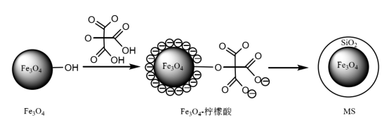
The preparation process of magnetic silicon based nanoparticles ( MS) is shown in Figure 1.
Figure 1 Schematic diagram of preparation of magnetic silicon-based nanoparticles
( 1) First, magnetic Fe3O4 nanoparticles modified with citric acid were prepared by chemical co-precipitation method, and the experimental procedure which comprises: 2.25 g of FeCl3·6H2O and 1.61 g of FeSO4·7H2O are added into three neck flask filled with 100 mL deionized water, the temperature of the system was heated to 85 ℃ under the condition of nitrogen gas . Stir under 1000 r·min-1 and quickly pour 10 mL of concentrated ammonia water. After the solution turns black, add 153 mg of sodium citrate. After 30 minutes of reaction, separate the production with a magnet, and wash the solid phase with deionized water and ethanol several times. The Fe3O4 nanoparticles obtained are dried for later use.
( 2) Then, magnetic silicon based nanoparticles (MS) were prepared by sol-gel method, shown in Figure 1. The experimental steps include: 0.1 g Fe3O4 nanoparticles were uniformly dispersed into a mixed solution containing 40 mL of anhydrous ethanol, 10 mL of dedistilled water and 1.2 mL of concentrated ammonia water. Under the stirring condition of 300 r·min-1, droplets of solution containing Fe3O4 nanoparticles were added to TEOS (0.4 mL) dispersed in 30 mL of anhydrous ethanol, and the reaction of silicon based nanoparticles was carried out for 12 h under the room temperature. The products were washed with anhydrous ethanol and distilled water for several times to obtain magnetic silica based nanoparticles (MS), which were dried for later use.
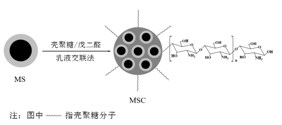
MSC microspheres were prepared by emulsion crosslinking method, as shown in Figure 2. The experimental steps included: 0.125 g magnetic silica nanoparticles (MS) evenly dispersed to 10 mL chitosan solution with mass fraction of 2.5% prepared by chitosan dissolved in acetic acid solution with volume fraction of 5.0%, and then add the chitosan solution into a emulsion containing 60 mL liquid paraffin wax and 1.8 mL twain 80 drop by drop. After stirring 30 min in the condition of 40 ℃ water bath and 800 r·min-1,add 2.0 mL glutaraldehyde and react 1 h. Then the pH of the system was adjusted to 9.0 with 1 mol·L-1 NaOH, and the reaction was continued for 1 h. The products were washed with petroleum ether, ethanol and distilled water for several times to obtain MSC microspheres, which were dried in vacuum for later use.
Figure 2 Schematic diagram of preparation of magnetic silicon based chitosan microspheres
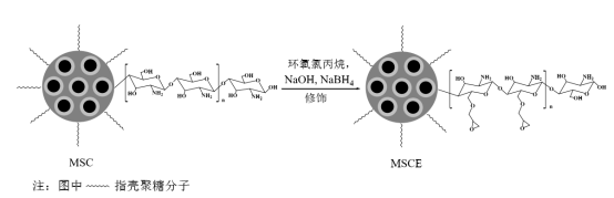
5. Preparation of epoxy based magnetic silicon based chitosan microspheres ( MSCE)
0.5 g of dried MSC microspheres prepared by the method of 4 were weighed and added into a conical flask of 50 mL. Then 2.0 mL of of NaOH solution, epichrolohydrin solution and NaBH4 solution were added into the flask with a certain concentration, respectively. And the reaction was carried out by constant temperature oscillation for a certain time. The reaction mechanism of preparation of MSCE is shown in Figure 3.
Figure 3 The principle of preparation of epoxy-based magnetic silicon-based chitosan microspheres
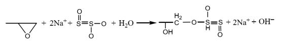
After activation, the activated microspheres are washed repeatedly with distilled water until there are no free OH- and epoxy groups on the surface to obtain epoxy based magnetic silicon based chitosan microspheres ( MSCE). The detection method is as follows: take 3.0 mL of the washing supernatant and add 1~2 drops of phenolphthalein. After shaking, if it didn’t turn red, it means there have no free OH- with MSCE; take 3.0 mL of the washing supernatant and add 3.0 mL of 1.3 mol·L-1 sodium thiosulfate and 1 ~2 drops of phenolphthalein, if the solution did not turning red after shaking, it means there have no free epoxy groups with MSCE. The reaction equation of the detection of epoxy group is as follows:
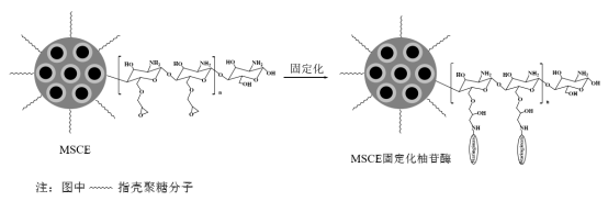
6 Preparation of MSCE immobilized naringinase enzyme
5.0 g of MSCE prepared by method 5 was added to 50 mL of naringinase enzyme solution with a certain pH, and the reaction was oscillated under a constant temperature water bath, as shown in Figure 4. After the reaction, the microspheres were washed with buffer solution for several times until no free naringinase was found on the surface of the microspheres. The microspheres were drained by a Brinella funnel to obtain MSCE immobilized naringinase enzyme, which was refrigerated at 4 ℃ for later use.
Figure 4 The principle of preparation of MSCE immobilized naringinase enzyme
Contact Us Now!
We accept customized services, we will usually contact you within 24 hours. You could also email me info@longchangadditive.com during working hours ( 8:30 am to 6:00 pm UTC+8 Mon.~Sat. ) or use the website live chat to get prompt reply.
This article was written by Longchang Chemical R&D Department. If you need to copy and reprint, please indicate the source.